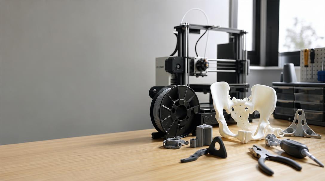

How a 3D-Printed Pelvis Model in Kuwait Changed Planning for Complex Hip Reconstruction
Planning a complex hip reconstruction may require more than simply examining images on a screen. At Jaber Al-Ahmad Hospital in Kuwait, a surgical team carried out a case involving the reconstruction of an artificial hip joint after preparing with a physical 3D-printed model of the patient’s pelvis. According to a VoxelMatters report dated September 5, 2026, this was the first case at the hospital to use this method.
This development shows that the value of 3D printing in the medical field extends beyond producing implants or prostheses. A physical copy of patient-specific anatomy can help the surgical team examine complex areas from different angles and discuss the preoperative approach in a more tangible way.
What was done at the hospital?
According to the report, the team used the patient’s imaging data to prepare a physical 3D model. This model was used as a planning tool before replacing the artificial hip joint and reassessing the surrounding bone structure. The critical point here is that the print itself was not the treatment; rather, the imaging data was transformed into an understandable, hands-on reference to support clinical decision-making.
The same production method or the same plastic may not be suitable for every case. If the model is intended to make bone geometry, critical gaps, and implant positioning visible, dimensional accuracy, surface detail, and rigidity appropriate to the model’s intended use should be evaluated together. For this reason, doctors, biomedical teams, and production specialists need to work together in in-hospital manufacturing processes.
Why is this news important for FDM users?
For Ucuz3D readers, the most practical takeaway is that custom parts should not be designed according to a mass-production mindset, but based on accurate data and a clearly defined purpose. In applications such as educational models, surgical rehearsal models, or anatomical communication mock-ups, FDM 3D printing can be an accessible option. However, an FDM-printed model should not be considered suitable for direct use as a medical implant; clinical implant production is a highly regulated, high-responsibility process with separate material, validation, sterilization, and regulatory requirements.
When making choices on the FDM side, the following questions come to the fore:
- Is the model intended only for visual and educational purposes, or is a higher level of detail required for surgical rehearsal?
- Will the part be transported frequently, or will it be a single-use presentation model?
- Is the surface generated from the imaging data suitable for printing and has it passed dimensional inspection?
- Do the model’s rigidity and color genuinely support the information expected in team communication?
The takeaway for Ucuz3D
Examples like this remind us that the value of 3D printing cannot be measured solely by material cost. A custom model prepared at the right time can facilitate communication, make planning meetings more tangible, and accelerate understanding of a complex part. At the same time, when anatomy, dimensions, and clinical decisions are involved, tolerances must be defined before production and checked after printing.
For a similar anatomical model, educational mock-up, or custom prototype, you can explore 3d printing service options. Understanding the price per gram model based on the part’s material and volume can help you carry out an initial feasibility assessment. If your file is ready, you can get a quick preliminary assessment using the online 3d printing tool. To clarify the material selection, also evaluate the options on the engineering materials 3D printing page together with your project’s operating conditions.
Points to consider in practice
In medical model printing, file preparation is at least as important as printer settings. Segmentation of imaging data, preservation of small anatomical details, and the choice of print orientation can affect the model’s usability. A large or complex model can be divided and printed in a controlled manner instead of being produced as a single piece; in that case, joining surfaces and reference markers should be designed from the outset.
In addition, dimensional inspection before and after printing should not be neglected, especially for models intended to support surgical planning. The fact that a model looks physically accurate does not automatically mean that every dimension has been verified. This approach positions the accessibility of FDM printing as a safe supporting tool that makes clinical decisions more understandable, without attempting to replace them.


 Türkçe
 العربية
 Русский
 فارسی
فارسی Deutsch